 |
Pigmentation
|
MODELS
PROTOCOL PRINCIPLE
The purpose of this study is to assess in vitro the pigmentation activity of the test product after systemic application (in the medium) on SkinEthicTM RHPE model for 6 days.
Ten reconstituted human tanned epidermal tissues (phototype IV) are dosed in the medium with the test product for 6 days. Half of the cultures (5 tissues) are also UVA/UVB irradiated. After the treatment, all tissues are scored visually. Additionally, one of the 5 tissues is assessed for histology (H&E staining) and one is used for tissue viability assessment. The remaining 3 tissues are used for melanin quantification.
DETAILED ASSAY PROCEDURE
Test method
Ten SkinEthicTM RHPE tissues (size 0.5 cm²) are treated (systemic application) with the test product and the positive control (a-MSH) for 6 days. Half of the cultures are irradiated with 50 mJ/cm² UVB / 1 J/cm² UVA, at day +1, +2 and +3 of the reception. After each dosing, all cultures (including fivefold untreated tissues) are consequently incubated at 37°C, 5% CO². Analysis (visual assessment, histology, MTT and melanin extraction) are performed after 6 days of treatment.
An evaluation of cell viability (MTT test) is performed.
At the end of the test period, one culture is placed on 300 µl of 0,5 mg/ml MTT and incubated 3 hours at 37° C 5% CO². Extraction is performed in 2 ml of isopropanol at room temperature, for a minimum of 2 hours. Optical density is measured at 570 nm. Results are expressed as percentage of viability compared to negative control: % Viability = [OD (570nm) tested compound / OD (570nm ) negative control)] x 100.
Histology
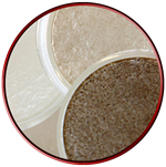
At the end of the test period, one culture is fixed in a balanced 10% formalin solution and embedded in paraffin. Four micron vertical sections are stained with hematoxylin/eosin and photographed under a microscope.
| not irradiated |
 |
| UVA/UVB irradiated |
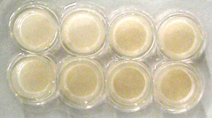
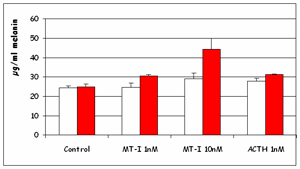
| The pigmentation is induced by UVA and UVB irradiation. The addition of MT-I and ACTH may provoke also an increase of pigmentation. These observations are confirmed by colorimetric melanin quantification performed after extraction with Soluene according to Ozeki protocol. |
 |
Visual assessment
At the end of the test period, all tissues (untreated, positive control and test material treated) are photographed using a digital camera for visual assessment of depigmentation.
Melanin extraction assay
At the end of the test period, for each condition, 3 tissues are removed from the insert by cutting out the filter with a sharp scalpel. The 3 filters are plunged into 360 µl of Solvable (Perkin Elmer) and heated at 100 degrees Celsius for 45 minutes. The optical density of the supernatants are measured on 80 µl extract at 490 nm using synthetic melanin as reference. Results are expressed as OD and as mg/ml melanin.
REFERENCES
Chemical characterisation of hair melanins in various coat-color mutants of mice. Ozeki, H., Ito S., Wakamatsu K. and Hirobe T., Journal of Investigative Dermatology, 105, 3, 361-366, 1995).
Human pigmented epidermis reconstructed in chemically defined medium used for evaluation of modulation of pigmentation. F. Sahuc, B. De Wever & M. Rosdy. Presented at the 12th Meeting of the European Society of Pigmented Cell Research, Paris, Sept. 2004.